
Mouse Secondary Antibodies

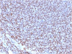
Biotium NKX2.2 (Neuroendocrine & Ewing's Sarcoma Marker)(NX2/2198R), CF647 conjugate, 0.1mg/mL
Expression of NKX2. 2 has been found in neuroendocrine tumors of the gut, making it a potential marker for the study of gastrointestinal neuroendocrine tumors. More recently, NKX2. 2 protein was identified as a target of EWS-FLI-1, the fusion protein specific to Ewing sarcoma, and was shown to be differentially upregulated in Ewing sarcoma on the basis of array-based gene expression analysis. It acts as a valuable marker for Ewing sarcoma, with a sensitivity of 93% and a specificity of 89%, and aids in the differential diagnosis of small round cell tumors. Primary antibodies are available purified, or with a selection of fluorescent CF Dyes and other labels. CF Dyes offer exceptional brightness and photostability. Note: Conjugates of blue fluorescent dyes like CF405S and CF405M are not recommended for detecting low abundance targets, because blue dyes have lower fluorescence and can give higher non-specific background than other dye colors.

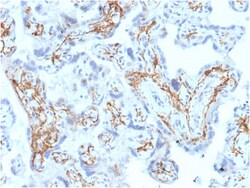
Biotium Glutathione S-Transferase Mu1 (GSTM1)(CPTC-GSTMu1-3), CF568 conjugate, 0.1mg/mL
Recognizes Glutathione S-Transferase of the mu class, which is involved in the detoxification of toxins, drugs, and products of oxidative stress by conjugation with glutathione. Primary antibodies are available purified, or with a selection of fluorescent CF Dyes and other labels. CF Dyes offer exceptional brightness and photostability. Note: Conjugates of blue fluorescent dyes like CF405S and CF405M are not recommended for detecting low abundance targets, because blue dyes have lower fluorescence and can give higher non-specific background than other dye colors.
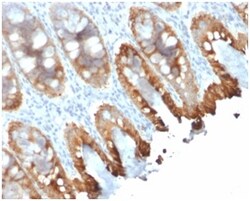
Biotium Cytokeratin 8/18(rKRT8.18/1346), 0.2mg/mL
Cytokeratin 8 (CK8) belongs to the type II (or B or basic) subfamily of high molecular weight cytokeratins and exists in combination with cytokeratin 18 (CK18). This MAb cocktail recognizes all simple epithelia including glandular epithelium, for example thyroid, female breast, gastrointestinal tract, respiratory tract, and urogenital tract including transitional epithelium. All adenocarcinomas and most squamous carcinomas are positive but keratinizing squamous carcinomas are usually negative. This antibody is useful in demonstrating the presence of Paget cells; there is very little keratin 18 in the normal epidermis so only Paget cells are stained. Immunohistochemical staining with this MAb is indistinguishable from that obtained with monoclonal antibody 5D3.Primary antibodies are available purified, or with a selection of fluorescent CF Dyes and other labels. CF Dyes offer exceptional brightness and photostability. Note: Conjugates of blue fluorescent dyes like CF405S an
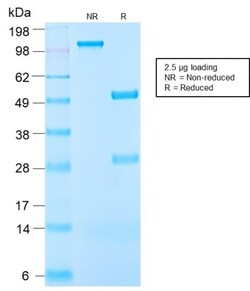
Biotium Insulin-like Growth Factor-1 (IGF-1) (IGF1/2872R), Biotin conjugate, 0.1mg/mL
This antibody is specific to Insulin-like Growth Factor (IGF-1) and shows minimal cross-reaction with IGF-11, Proinsulin, MSF, and Insulin. IGF-1 is a polypeptide growth factor with two isoforms that are produced by alternative splicing. Isoform 1 is also known as IGF-IB while isoform 2 is known as IGF-IA. IGF-1 stimulates the proliferation of a wide range of cell types including muscle, bone and cartilage tissue. It functions as an autocrine regulator of growth. Activation of IGF system has emerged as a key factor for tumor progression and resistance to apoptosis in many cancers like those of breast, thyroid and colon.Primary antibodies are available purified, or with a selection of fluorescent CF Dyes and other labels. CF Dyes offer exceptional brightness and photostability. Note: Conjugates of blue fluorescent dyes like CF405S and CF405M are not recommended for detecting low abundance targets, because blue dyes have lower fluorescence and can give higher non-specifi
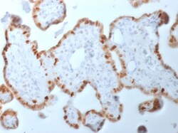
Biotium DNMT1 / DNA Methyltransferase 1(DNMT1/2061), 0.2mg/mL
This gene encodes an enzyme that transfers methyl groups to cytosine nucleotides of genomic DNA. This protein is the major enzyme responsible for maintaining methylation patterns following DNA replication and shows a preference for hemi-methylated DNA. Methylation of DNA is an important component of mammalian epigenetic gene regulation. Aberrant methylation patterns are found in human tumors and associated with developmental abnormalities. Variation in this gene has been associated with cerebellar ataxia, deafness, and narcolepsy, and neuropathy, hereditary sensory, type IE. Primary antibodies are available purified, or with a selection of fluorescent CF Dyes and other labels. CF Dyes offer exceptional brightness and photostability. Note: Conjugates of blue fluorescent dyes like CF405S and CF405M are not recommended for detecting low abundance targets, because blue dyes have lower fluorescence and can give higher non-specific background than other dye colors.
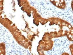
Biotium Villin (GI-Mucosal & Urogenital Brush Border Marker)(VIL1/2376), 0.2mg/mL
Recognizes a protein of 95 kDa, which is identified as villin. It is a major constituent in the microvilli, which compose the brush border of epithelial cells forming absorptive surfaces of the intestinal and renal proximal tubular epithelia. Anti-Villin labels the brush border area in the gastrointestinal mucosal epithelium and urogenital tract. Among neoplasms, villin is predominantly expressed in tumors of colorectal origin. Antibody to villin is useful in identifying malignant cells from primary and metastatic colorectal carcinomas. This antibody also labels Merkel cells of the skin. Primary antibodies are available purified, or with a selection of fluorescent CF Dyes and other labels. CF Dyes offer exceptional brightness and photostability. Note: Conjugates of blue fluorescent dyes like CF405S and CF405M are not recommended for detecting low abundance targets, because blue dyes have lower fluorescence and can give higher non-specific background than other dye colors.
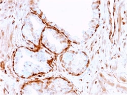
Biotium Aldo-keto Reductase Family 1 Member B1 (Adrenal Marker)(CPTC-AKR1B1-3), 0.2mg/mL
Aldo-keto Reductase Family 1 Member B1 (AKR1B1), also known as aldose reductase or aldehyde reductase, is a member of the aldo/keto reductase superfamily, which consists of more than 40 known enzymes and proteins. This protein catalyzes the reduction of a number of aldehydes, including the aldehyde form of glucose, and is thereby implicated in the development of diabetic complications by catalyzing the reduction of glucose to sorbitol. It has also been shown to have decreased expression in adrenocortical cancer, and possibly play a role in adrenal tumorigenesis. It has been suggested that AKR1B1 could be investigated as a marker of malignancy for adrenal tumor diagnosis.Primary antibodies are available purified, or with a selection of fluorescent CF Dyes and other labels. CF Dyes offer exceptional brightness and photostability. Note: Conjugates of blue fluorescent dyes like CF405S and CF405M are not recommended for detecting low abundance targets, because blue dyes have lower
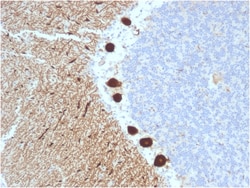
Biotium Carbonic Anhydrase VIII(CPTC-CA8-2), CF568 conjugate, 0.1mg/mL
Carbonic anhydrase VIII (carbonic anhydrase 8) was named because of sequence similarity to other known carbonic anhydrase genes. However, the gene product lacks carbonic anhydrase activity (i. e. , the reversible hydration of carbon dioxide). The gene product continues to carry a carbonic anhydrase designation based on clear sequence identity to other members of the carbonic anhydrase gene family. Mutations in the gene cause neurological disorders in mice. Primary antibodies are available purified, or with a selection of fluorescent CF Dyes and other labels. CF Dyes offer exceptional brightness and photostability. Note: Conjugates of blue fluorescent dyes like CF405S and CF405M are not recommended for detecting low abundance targets, because blue dyes have lower fluorescence and can give higher non-specific background than other dye colors.
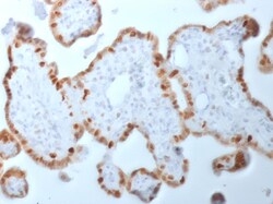
Biotium DNMT1 / DNA Methyltransferase 1(DNMT1/2061), 1mg/mL
This gene encodes an enzyme that transfers methyl groups to cytosine nucleotides of genomic DNA. This protein is the major enzyme responsible for maintaining methylation patterns following DNA replication and shows a preference for hemi-methylated DNA. Methylation of DNA is an important component of mammalian epigenetic gene regulation. Aberrant methylation patterns are found in human tumors and associated with developmental abnormalities. Variation in this gene has been associated with cerebellar ataxia, deafness, and narcolepsy, and neuropathy, hereditary sensory, type IE. Primary antibodies are available purified, or with a selection of fluorescent CF Dyes and other labels. CF Dyes offer exceptional brightness and photostability. Note: Conjugates of blue fluorescent dyes like CF405S and CF405M are not recommended for detecting low abundance targets, because blue dyes have lower fluorescence and can give higher non-specific background than other dye colors.
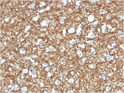
Biotium p14ARF(4C6/4), 1mg/mL
The progression of cells through the cell cycle is regulated by a family of proteins designated cyclin-dependent kinases (Cdks). Sequential activation of individual members of this family and their consequent phosphorylation of critical substrates promotes orderly progression through the cell cycle. Multiple proteins are encoded by the tumor suppressor gene CDKN2A (MTS1/ p16INK4a) via translation of alternate reading frames, resulting in the pro- duction of the p19 ARF protein in mice and the p14 ARF protein in humans. p14 ARF induces an increase in MDM2 and p21 levels and leads to cell cycle arrest in both G1 and G2/M. p14 ARF is negatively regulated by p53 and is known to bind directly to MDM2. CDKN2A also encodes the mitotic protein p16, which binds to and inhibits the Cdk4/cyclin D complex. Primary antibodies are available purified, or with a selection of fluorescent CF Dyes and other labels. CF Dyes offer exceptional brightness and photostability. Note: Conjugates of blue

Biotium FABP2 (Marker of Metastatic Potential in Colorectal Cancer)(CPTC-FABP2-3), CF488A conjugate, 0.1mg/mL
The intracellular fatty acid-binding proteins (FABPs) belong to a multigene family with nearly twenty identified members. FABPs are divided into at least three distinct types, namely the hepatic-, intestinal- and cardiac-type. They form 14-15 kDa proteins and are thought to participate in the uptake, intracellular metabolism and/or transport of long-chain fatty acids. They may also be responsible in the modulation of cell growth and proliferation. Intestinal fatty acid-binding protein 2 gene contains four exons and is an abundant cytosolic protein in small intestine epithelial cells. This gene has a polymorphism at codon 54 that identified an alanine-encoding allele and a threonine-encoding allele. Thr-54 protein is associated with increased fat oxidation and insulin resistance. Primary antibodies are available purified, or with a selection of fluorescent CF Dyes and other labels. CF Dyes offer exceptional brightness and photostability. Note: Conjugates of blue fluorescent dyes
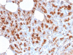
Biotium MDM2(MDM2/2414), CF488A conjugate, 0.1mg/mL
MDM2 is a nuclear phosphoprotein that binds and inhibits transactivation by tumor protein p53. It can promote tumor formation by targeting tumor suppressor proteins, such as p53, for proteasomal degradation. Overexpression of MDM2 can result in excessive inactivation of tumor protein p53, diminishing its tumor suppressor function. This protein also affects the cell cycle, apoptosis, and tumorigenesis through interactions with other proteins, including retinoblastoma 1 and ribosomal protein L5. Overexpression of MDM2 protein is detected in a variety of cancers. Primary antibodies are available purified, or with a selection of fluorescent CF Dyes and other labels. CF Dyes offer exceptional brightness and photostability. Note: Conjugates of blue fluorescent dyes like CF405S and CF405M are not recommended for detecting low abundance targets, because blue dyes have lower fluorescence and can give higher non-specific background than other dye colors.

Biotium Myoglobin (Muscle Cell Marker)(MB/2105), 0.2mg/mL
Myoglobin is a cytosolic oxygen-binding protein responsible for the storage and diffusion of oxygen within myocytes. Expression of myoglobin is highest in skeletal and cardiac muscle. Myoglobin is well accepted as an O2-storage protein in muscle, capable of releasing O2 during periods of hypoxia or anoxia. In combination with other striated muscle markers such as vimentin and myogenin, myoglobin is helpful in the identification of rhabdomyosarcoma and tumors with skeletal muscle differentiation. Reportedly, myoglobin is expressed on epithelial cancer cells due to changed metabolic and environmental conditions. Primary antibodies are available purified, or with a selection of fluorescent CF Dyes and other labels. CF Dyes offer exceptional brightness and photostability. Note: Conjugates of blue fluorescent dyes like CF405S and CF405M are not recommended for detecting low abundance targets, because blue dyes have lower fluorescence and can give higher non-specific background tha
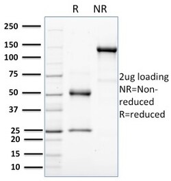
Biotium HER-4 / ERBB4(HFR-1), 1mg/mL
The EGF receptor family comprises several related receptor tyrosine kinases that are frequently overexpressed in a variety of carcinomas. Members of this receptor family include EGFR (HER1), Neu (ErbB-2, HER2), ErbB-3 (HER3) and ErbB-4 (HER4), which form either homodimers or heterodimers upon ligand binding. The gene encoding ErbB-4 is expressed as a full-length protein, which produces a short membrane-anchored cytoplasmic domain fragment and a long ectodomain fragment. The short fragment is heavily tyrosine phosphorylated and possesses tyrosine kinase catalytic activity toward an exogenous substrate. Proteolytic cleavage of ErbB-4 is promoted by the binding of heregulin. ErbB-4 is involved in cell proliferation and differentiation and its expression is highest in breast carcinoma cell lines, normal skeletal muscle, heart, pituitary, brain and cerebellum. Its expression in breast cancer, pediatric brain cancer and other types of carcinomas has been reported in studies which sugges
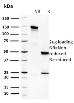
Biotium Spermidine or Spermine N1-Acetyltransferase 1(CPTC-SAT1-3), 1mg/mL
Spermidine/spermine N1-acetyltransferase 1 (SAT1 or SSAT1) is the key regulatory enzyme in the catabolism of polyamines, catalyzing acetylation of spermidine or spermine to generate N1-acetyl spermidine or N1-acetyl spermine, and N1, N12-diacetylspermine. The cellular level of SAT1 is normally extremely low, but it is induced rapidly by a variety of stimuli, including polyamines, polyamine analogs, toxic chemicals, certain drugs, and growth factors. Downregulation of SAT1 has been reported in Epstein-Barr virus positive Burkitt's lymphoma cells. Primary antibodies are available purified, or with a selection of fluorescent CF Dyes and other labels. CF Dyes offer exceptional brightness and photostability. Note: Conjugates of blue fluorescent dyes like CF405S and CF405M are not recommended for detecting low abundance targets, because blue dyes have lower fluorescence and can give higher non-specific background than other dye colors.